Cell Imaging Service (CICS)
About us
The Centre Imagerie Cellulaire Santé (CICS) offers three complementary technical platforms to research lab and industrials:
- electron microscopy (TEM and SEM) + sample preparation
- photon microscopy (MP) + sample preparation
- Flow cytometry (CMF)
Our services
- Five full-time staff
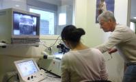
- Full support from sample preparation to image interpretation
- Training in the use of imaging equipment and sample preparation
- Scientific and technical watch
- Our main themes = Animal / Plant Biology, Health, Pharmaceuticals / Cosmetics, Environment, Nutrition / Agrifood, Biomaterials, Nanotechnologies, Metallurgy, etc.
Facilities / Equipments
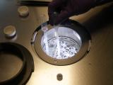
Located on 330m² on the ground floor of UFR Medicine, the CICS offers 3 technical rooms equipped with automated sample preparers and imaging equipments:
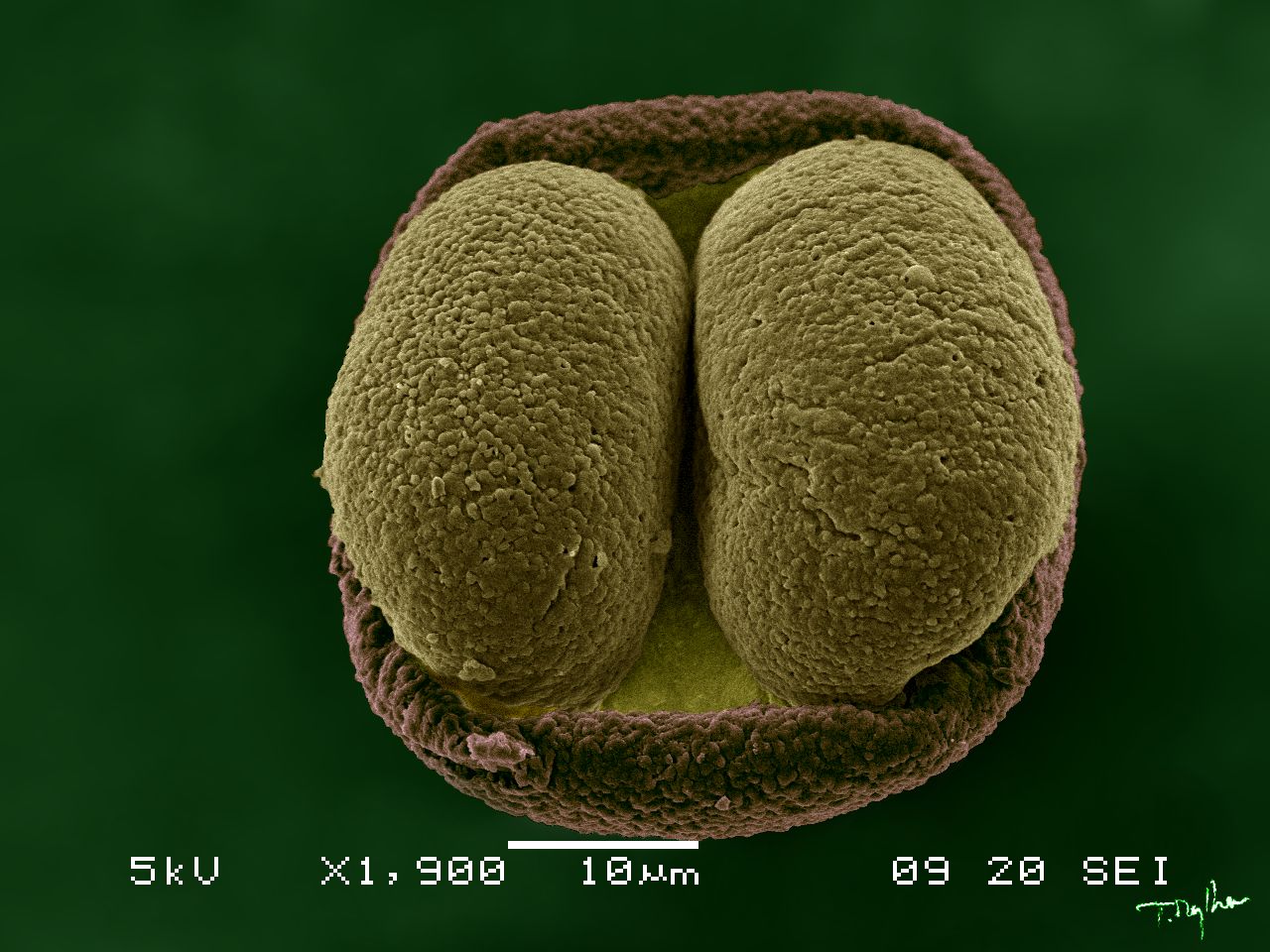
- Hitachi H7650 Transmission Electron Microscope + Hamamatsu Camera
- Jeol 6060LV Scanning Electron Microscope
- BD LSR II Flow Cytometer
- Olympus BX51 Light-field Microscope + Color View Camera
- Olympus BX51 Fluorescence Microscope + CoolSnapES Camera
Some of our achievements
- 1,2,3‐Triazolium‐Based Cationic Amphipathic Peptoid Oligomers Mimicking Antimicrobial Helical Peptides. Dr. Radhe Shyam, Nicolas Charbonnel, Aurélie Job, Christelle Blavignac, Christiane Forestier, Claude Taillefumier, Sophie Faure. ChemMedChem. 19 June 2018.
- Development and cytotoxic response of two proliferative MDA-MB-231 and non-proliferative SUM1315 three-dimensional cell culture models of triple-negative basal-like breast cancer cell lines. Dubois Clémence, Dufour Robin, Daumar Pierre, Aubel Corinne, Szczepaniak Claire, Blavignac Christelle, Mounetou Emmanuelle, Penault-Llorca Frédérique and Mahchid Bamdad. Oncotarget 2017.
- Absence of Fungal Spore Internalization by Bronchial Epithelium in Mouse Models Evidenced by a New Bioimaging Approach and Transmission Electronic Microscopy. Am J Pathol. 2015. Rammaert B, Jouvion G, de Chaumont F, Garcia-Hermoso D, Szczepaniak C, Renaudat C, Olivo-Marin JC, Chrétien F, Dromer F, Bretagne S.
- Roles of capsule and lipopolysaccharide O antigen in interactions of human monocyte-derived dendritic cells and Klebsiella pneumoniae. Evrard B, Balestrino D, Dosgilbert A, Bouya-Gachancard JL, Charbonnel N, Forestier C, Tridon A. Infect Immun. 2010.
Training
- Training in the use of imaging equipment and sample preparation
- Training in different diploma
Contacts
Administrative & scientific leader
Tel.: +33 (0)4 73 17 80 88
Technical manager
Flow cytometry-SEM expert
Tel.: +33 (0)4 73 17 80 88
TEM expert
Tel.: +33 (0)4 73 17 80 87
Photon microscopy expert
Tel.: +33 (0)4 73 17 80 86
Access to the service
CICS is open from 8 am to 5 pm from Monday to Friday without any geographical, thematic or institutional restrictions. The appointments are by email: cics@uca.fr or by phone +33 (0) 4 73 17 80 88.
Address
Centre Imagerie Cellulaire Santé
UFR Médecine
28 place Henri Dunant
63000 Clermont-Ferrand
Bâtiment principal
France
UFR Médecine
28 place Henri Dunant
63000 Clermont-Ferrand
Bâtiment principal
France