Microprobe Scanning Electron Microscopy (EPMA)
About us
 The SEM (JEOL 5910 LV) is optimized for secondary and backscattered electron imaging, it is also equipped with a cathodoluminescence detector. It also makes it possible to carry out analyzes by energy dispersive spectrometry (EDS).
It can operate in controlled vacuum for specific needs, without the metallization of vacuum-sensitive samples.
The EDS micro-analysis system is a Bruker brand SDD detector. The system manages qualitative and semi-quantitative chemical analyzes, acquisition of spectra, concentration profiles and chemical mappings. It can be coupled to an EBSD detector (Bruker) for the acquisition of orientation and phase recognition mappings.
Our services
The electron microscopy service is dedicated to the preparation, imaging and analysis of inorganic samples. It is mainly used by different university laboratories, but can be opened to private companies.
The service is also equipped with a Denton metallizer for carbon evaporation and Gold-Palladium metallization (Au-Pd) samples.
The service is also equipped with a Denton metallizer for carbon evaporation and Gold-Palladium metallization (Au-Pd) samples.
Equipments
The SEM uses an electron beam, emitted by an electron gun. The interaction between the electrons and the sample generates many phenomena.
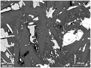
The secondary electrons (SE) allow imaging of the sample topography. 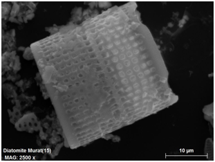 The backscattered electrons (BSE) generates a "chemical" contrast image according to the atomic mass of the phases present in the sample, so a bright phase will reveal an object composed of heavy chemical elements.
The secondary electrons (SE) allow imaging of the sample topography.

Diatomée - Echantillon Collection de Minéralogie-LMV
Image en électrons secondaires - J. Marc HENOT – LMV

Oxyde de fer dans un basalte provenant de Chine
Image en électrons rétrodiffusés - J. Marc HENOT – LMV
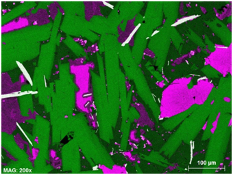
With the EDS detector which will capture the energy of the X-rays emitted during the interaction of the atoms present in the sample with that of the beam emitted by the gun, it is possible to know the chemical composition of the latter. This analysis is qualitative and semi-quantitative, it may be punctual by spectral analysis or characterize a surface by mapping by assigning a color to each chemical element.

Cartographie d’un basalte provenant de Chine
Juxtaposition de trois cartographies mono élémentaire : Vert=Al, Rouge=Fe, Bleu=Ti - J. Marc HENOT - LMV
The EBSD detector uses the diffraction of backscattered electrons and the acquisition of the maps restores the recognition, the distribution of the phases and their crystalline orientations. The Quantax system (Bruker) allows the simultaneous acquisition of chemical and orientation maps. The polishing of the samples is particular for this technique.
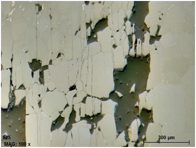
Forsterite – Echantillon D. Freitas
Image combinée FSE-BSE
J. Marc HENOT – LMV
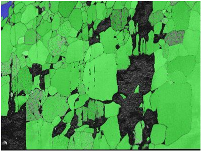
Cartographie de phases Forsterite (vert)
Pyrope (Bleu)
image J. Marc HENOT - LMV
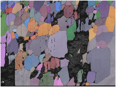
Cartographie de distribution des grains
dit de Euler
Image J. Marc HENOT - LMV
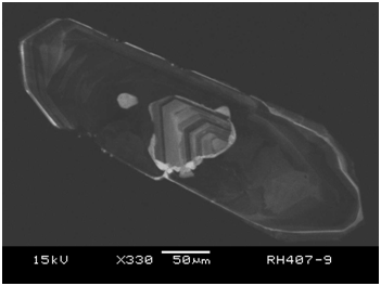
Under the impact of the electron beam the cathodolumiscence, allows to visualize for some oxides the variation of structure or composition, like zonations of growth.

Zonations de croissance d’un cristal d’oxyde de zirconium – Echantillon V. Bosse
Image en cathodolumiscence - J. Marc HENOT - LMV
Image en cathodolumiscence - J. Marc HENOT - LMV

Contact
Technical expert
Address
Laboratoire Magmas et Volcans
Campus Universitaire des Cézeaux
6 Avenue Blaise Pascal
Campus Universitaire des Cézeaux
6 Avenue Blaise Pascal
63178 AUBIERE Cedex
France

